We discussed coloboma disease in our previous blog about coloboma renal, and now let’s get into understanding what ocular coloboma is, or, we can say, eye coloboma syndrome. Apart from chorioretinal coloboma, there are five distinct types of coloboma which are discussed in this article.
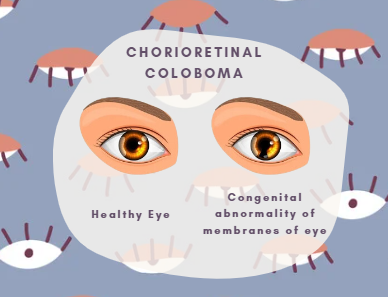
Eye coloboma syndrome, also termas ocular coloboma, is a genetic condition in which specific eye tissues either fail to develop at the fetal stage or are wholly absent at the time of birth, which results in congenital vision impairment. Ocular coloboma may affect one eye (unilateral coloboma eye) or both eyes (bilateral coloboma eyes). However, some cases are with completely non-functional eyes, while some others recognize with minor structural and functional eye defects.
Usually, Coloboma eyes can be visibly detected by examining the iris. In a typical coloboma iris, a small and underdevelop iris, called a hypoplastic iris, is evident. In the examination of the pupil, the patient has a keyhole-shaped pupil known as a keyhole eye/keyhole pupil. Remember, another term for such coloboma eyes is teardrop pupil/teardrop-shaped pupil.
Types of Ocular Coloboma
Generally, based on location, there are six eminent types of coloboma eyes. These types are discuss hereunder.
1. Chorioretinal coloboma.
As the name indicates, this type of coloboma mainly targets the retina (the part of the eye that detects light). In this type of coloboma eye, a layer of cells in the retina is missing, which leads to a gap between the retina and the choroid layer of the eye. Hence it is also known as choroidal coloboma.
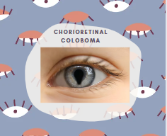
Hence, the retina fails to detect light and sends impaired signals to the optic nerve, which directly affects the patient’s vision. Retinal detachment and reduced visual acuity are two visual impairments associated with retinal coloboma.
Coloboma retina usually occurs due to the failure of the embryonic fissure to close during development.
Clinical Features of Chorioretinal Coloboma.
The significant signs of chorioretinal coloboma are;
- The posterior globe is mainly affect by retinal coloboma.
- The Coloboma retina is usually located in the inferotemporal area.
- It is characterizes as both unilateral and bilateral coloboma.
- Chorioretinal coloboma is generally link with other coloboma eye disorders, including the iris or optic nerve.
- At the margin of coloboma eyes, choroidal neovascularization may occur.
2. Eyelid Coloboma.
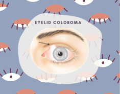
In this type of coloboma eyes, either a complete eyelid or a part of it is missing. Coloboma of the eyelid is mainly due to the lack or underdevelopment of eyelid tissues, which occurs at the junction of the superior medial and middle third of the upper eyelid. Apart from phenotypic appearance, it can affect vision and the cornea.
Eyelid coloboma may result from other coloboma-associated syndromes, such as Goldenhar syndrome, Treacher-Collins syndrome, and CHARGE syndrome. One of the closely associated syndromes is Manitoba Oculotrichoanal (MOTA), characterized by unilateral upper eyelid coloboma and bifid nose.
Clinical Features of Eyelid Coloboma.
- The most common features of eyelid coloboma are dermal scarring and defective orbicularis muscles.
- Eyelid coloboma is a full-thickness defect of the eyelid.
- The coloboma of the eyelid is usually bilateral or sometimes unilateral.
- A partial or complete absence of an eyelid characterizes it.
- Eyelid coloboma involves defects like a shortage of conjunctiva, tarsal plate, orbicularis muscle, and skin.
3. Lens Coloboma.
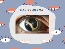
In this type of ocular coloboma, the lens (the part of the eye that resides behind the iris and focuses light on the retina) is missing, or the equator of the lens is flattened. Hence, light will not focus on the retina, and no signals are receive by the optic nerve, which results in vision impairment.
The primary cause of lens coloboma is the absence or faulty development of zonular fibers and ciliary bodies (that hold the lens in place). In such coloboma eyes, the lens is usually deprive of its standard pull in the defective region, which is thicker and somewhat funny-shaped. Likewise, in other ocular colobomas, lens coloboma is unilateral or bilateral.
4. Macular Coloboma (Sub Class of Chorioretinal Coloboma).
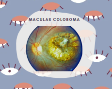
In macular coloboma, the macular (the central portion of the retina that focuses on details by giving sharp, central vision) fails to develop or is completely missing. It occurs due to the failure of fusion of the optic fissure during developmental stages. It is usually a unilateral coloboma.
Macular coloboma is characterizes by a sharply defined, rather large defect in the central area of the fundus that is well circumscribed, oval or round, atrophic lesions of varying size presenting rudimentary or absent retina and coarsely pigmented.
5. Optic Nerve Coloboma.
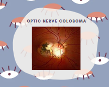
It is eminently known as coloboma of the optic disc. In optic nerve coloboma, the optic nerve that sends light signals from the eye to the brain is partially develop or hollow from the inside, which results in moderate to severely reduced vision. This defect involves the inferiorly located optic nerves present in the optic disc and happens due to incomplete closure of embryonic fissures in pregnancy.
Clinical Features of Optic Nerve Coloboma.
- Optic nerve coloboma is usually a unilateral or bilateral coloboma disorder.
- It may be present sporadically or inherited in an autosomal dominant fashion.
- Optic nerve coloboma involves visual field defects and relative afferent pupillary defects (RAPD).
- It is mainly characterize by glimmering white, circumscribes sharply, enlarged, and deeply hollowed out optic disc.
- A person with coloboma of the optic disc may have a macular or retinal detachment.
- Microcornea (small cornea), retinal dysplasia, and microphthalmos (small eyes) are common symptoms of optic nerve coloboma.
- Optic nerve coloboma may result from other coloboma-associated syndromes, such as Meckel’s syndrome, Aicardi’s syndrome, and CHARGE syndrome.
6. Uveal Coloboma.
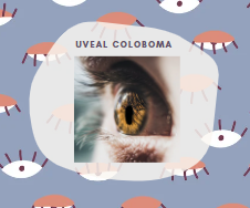
In this type of coloboma, the uvea is missing. It is the central part of the eye that connects with the iris, choroid, and ciliary body, thus hindering the proper detection of light. Moreover, uveal coloboma is the foremost reason for childhood blindness due to missing ocular tissues.
Cause of Uveal Coloboma.
The primary cause of uveal coloboma is the defective closure or even failure of closure of embryonic optical fissure at a gestational age of five to seven weeks.
What Causes Ocular Coloboma?
As we know, coloboma is a mutilated condition so, just like renal coloboma syndrome, eye coloboma syndrome has the same genetic basis, i.e., a mutation in a gene called PAX-2 gene. PAX-2 gene primarily encodes proteins that are responsible for the healthy development of eyes, ears, brain, and kidneys. This underdevelopment leads to the malfunctioning of respective organs. However, the fact that the kidneys and eyes are majorly target by the mutation is still unclear. Here, it must be notice that certain environmental factors, like having alcohol during pregnancy, may also cause an elevation in the risk of ocular coloboma.
An interesting fact is the yield of coloboma cases via molecular genetic testing is relatively low, which suggests that environmental conditions majorly contribute to the etiology of this condition.
Symptoms of Ocular Coloboma.

Some commonly detected signs of ocular coloboma include the following.
- Low or blurry vision.
- Complete blindness.
- Inability to focus correctly.
- Sensitivity to light.
- Failure to control eye movements.
- More significant than the usual blind spot.
- Keyhole-shaped pupil.
Treatment of Chorioretinal Coloboma.
Now, indeed, there is no way to produce new tissues or replace the missing eye part in case of ocular coloboma. This means that focus on prevention is more necessary than treatment. However, some management can be made to improve the quality of a patient’s life. These management measures are depicted below:
- People with lenses and iris coloboma can use glasses with corrected lenses and colored lenses to have corrected vision. This management will be lifelong.
- Surgeries can be perform to correct eyelid coloboma and sometimes to make the pupil look rounder so that the correct amount of light can be fetch and focus by the patient.
- Suppose the vision is completely lost and can’t be corrected by glasses. Likewise, in such case, visual aids like magnifiers and screen readers are recommended to the patient to improve their quality of life.
NOTE: Coloboma is genetic, and it is not your fault if you suffer from the respective condition. Instead of worrying, focus on the management to have a better life.
